Laboratory examinations have been used by doctors for centuries to confirm disease diagnoses. Before the discovery of the microscope, laboratory examinations were conducted macroscopically, relying on the bare eyes to observe various types of specimens such as urine and feces. However, macroscopic examinations alone could not reveal microscopic changes, such as the type and form of parasitic cells, bacteria, and viruses. In 1590, Zacharias Janssen and Hans Janssen, a son and a father, invented the light microscope, which enabled the magnification of small objects that were otherwise invisible to the bare eyes. This invention paved the way for new discoveries and research in the fields of biology, medicine, and other sciences. As time goes by, laboratory examination methods for detecting and measuring target substances in samples have been evolving. Examples of these advancements are the RID (Radial immuno diffusion) and RIA (Radio immunoassay) technologies.
In the development of microscopic laboratory examination technology, the ELISA (enzyme-linked immunosorbent assay) method has become a significant breakthrough. This technology was discovered by Eva Engvall and Peter Perlman in 1971. The ELISA method is applied in various fields and has become a routine technique used in research laboratories and diagnostics worldwide. They modified the radioimmunoassay (RIA) method by replacing the radioactive iodine 125 with an enzyme to conjugate antigens and antibodies.
The ELISA method was first used to determine the level of IgG antibodies in rabbit blood serum. The ELISA method was eventually patented in the United States and Europe. After that, the ELISA method began to be widely used by researchers. In 1974, LJungstrom et al. utilized this method in parasitology to identify trichinosis. In 1945, Voller used the ELISA method to diagnose malaria. From 1978 to 1981, Bishai, Galli, Leinikki, Ukkonen, et al. used the ELISA method to identify infections caused by influenza, parainfluenza, and mumps viruses. In 1980, Siegle et al. modified the ELISA method with microtiter plates to identify the concentration of various hormones, peptides, and proteins. Currently, the ELISA method is widely used for research and diagnostic purposes such as COVID virus antigen testing, hepatitis screening, parasite analysis, tumor marker detection, hormone level measurement, and others.
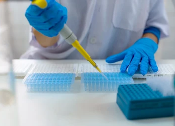
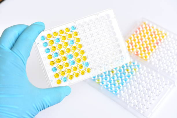
ELISA operates on two principles: the reaction between antibodies and antigens, and the reaction between enzymes and substrates. These reactions occur in each microplate well. The well surface is coated with specific antibodies against the target antigen. When a sample containing the target antigen is added to the well, a reaction occurs between the antibodies and the antigen, forming an antibody-antigen bond. However, the sample may contain other unwanted antigens, which are eliminated by washing with a buffer. The antibody-antigen complexes are then bound by antibodies labeled with enzymes (enzyme-labeled antibodies). To determine the presence of an antibody-antigen bond, a substrate is added, which reacts with the enzyme on the enzyme-labeled antibody, resulting in the formation of a colored product. To stop this reaction, NaOH, HCl, or sulfuric acid is added. The color formed is measured for absorbance at a wavelength of 400-600 nm using an ELISA reader based on the principles of spectrophotometry.
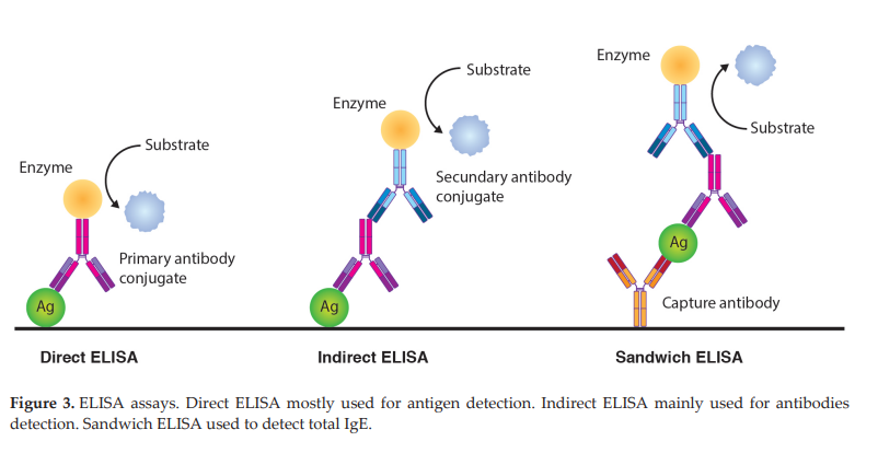
There are three types of ELISA methods:
- Direct ELISA (antigen screening)
In direct ELISA, the antigen is directly bound to enzyme-labeled antibodies. This method is commonly used to measure antigen concentrations. - Indirect ELISA
In indirect ELISA, the antigen is not directly bound to enzyme-labeled antibodies but is first bound to primary antibodies. This method is commonly used to measure total antibodies in a sample. - Sandwich ELISA (antibody screening): Unlike direct and indirect ELISA, the sandwich ELISA involves coating the well with capture antibodies. These antibodies bind to the target antigen and are subsequently bound by direct and indirect antibodies, forming a sandwich-like structure. This method is commonly used to measure the antigen concentration in complex samples, such as hormones and cytokines.
This method has advantages such as:
- Simple procedure
- High specificity and sensitivity, as it is based on the specific reaction between antigens and antibodies
- High efficiency
- Generally safe and environmentally friendly, as it does not require radioactive substances and large amounts of organic solvents
However, ELISA also has disadvantages such as:
- High cost of antibody preparation
- Requires complex techniques and expensive culture media
- Possibility of high false-positive/false-negative results
- Antibody instability
- Requires transportation and storage in a cold condition due to the instability of antibodies
In conclusion, laboratory examinations have undergone significant evolution since the discovery of the light microscope by Zacharias Janssen and Hans Janssen in 1590. The development of the ELISA (enzyme-linked immunosorbent assay) method by Eva Engvall and Peter Perlman in 1971 marked an important milestone in microscopic laboratory testing. This method has made significant contributions to disease diagnosis, biological research, medicine, and other scientific fields. In the ELISA method, the reaction between antigens, antibodies, and enzymes with substrates occurs within a well microplate, and the results can be measured through spectrophotometry. ELISA has advantages such as simple procedures, high sensitivity, and good efficiency. However, there are some challenges, such as the high cost of antibody preparation and the possibility of false positive/negative results. Nevertheless, ELISA remains one of the most commonly used methods in laboratory examinations today, with various applications in disease diagnosis, health monitoring, and research. With the continuous advancement of technology, it is expected that the ELISA method will continue to evolve and make even greater contributions to improving healthcare quality and scientific understanding in the future.
References:
American Physical Society.Lens Crafters Circa 1590: Invention of the Microscope. APS News. 2004. https://www.aps.org/publications/apsnews/200403/history.cfm
Aydin S. A short history, principles, and types of ELISA, and our laboratory experience with peptide/protein analyses using ELISA.Peptides. 2015;72:4-15.
Sakamoto S, Putalun W, Vimolmangkang S, Phoolcharoen W, Shoyama Y, Tanaka H, Morimoto S. Enzyme-linked immunosorbent assay for the quantitative/qualitative analysis of plant secondary metabolites. J Nat Med. 2018;72(1):32-42. doi: 10.1007/s11418-017-1144-z
Leave a Reply